Installation
Run DrawWing-0.45_setup and follow the instructions.
Browse wing images
Use Menu File > Open (Ctrl+O) to view single file. DrawWing opens images in following formats: PNG, BMP, JPG, TIF. I prefer PNG because of its good compression without loss of quality. If the information about resolution is not included in the image file user needs to provide this information. Without correct information about resolution DrawWing will not work correctly. In case of standard wing images (with extension .dw.png) information about measurement points will be displayed as well. It is possible to correct the position of the points using drag-and-drop method. The changes of the points position need to be saved using Menu File > Save data (Ctrl-S). In order to obtain coordinates of the points use Menu View > Data (Ctrl+D). The coordinates are in bitmap coordinate system - (0,0) point is in the upper left corner of the image. Coordinates of the vein junctions in Cartesian coordinate system can be saved in TPS format using Menu File > Export to TPS. The TPS files can be used with TPS software. Indices used by beekeepers can be obtained using Menu View > Apis indices. The indexes can be further analysed using MorphPlot by Peter Edwards. In order to brows larger number of standard wing images use Menu File > Open dw.png directory. In order to navigate between images from the folder use Menu File > Open next file (Ctrl+Right) or Open previous file (Ctrl+Left).
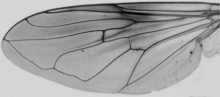
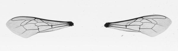 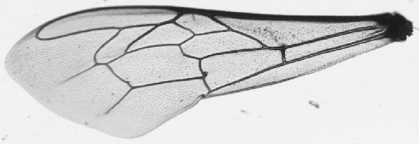 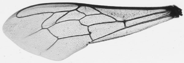 ____________________ ____________________Analyse wing images In order to analyse single wing image displayed in DrawWing window use Menu Wing > Apis junctions (Ctrl+A). If the image is large and contains many images the analysis can take relatively long time. Progress bar displayed at the status bar can be used to estimate the remaining time of the analysis. In order to analyse many wing image from one folder use Menu Wing > Apis junctions dir. During analysis wings detected in an image are cropped out and saved as Standard wing images in the same folder as the analysed image. The file names of the created images are the same of the analysed image but with appended wing number and information if this is a right (R) or left wing (L). In case of the image shown here two standard wing images were created. Smaller hind wings at the moment are not detected by DrawWing. You can use "Step by step" option in order to control more precisely the analysis. In step by step analysis of images with more than one wing use Menu Wing > Next step. |
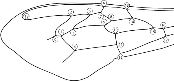 ____________________ ____________________Classification of honeybee (Apis mellifera) subspecies Before you can perform the classification you need to analyse a set of forewings from one colony. The standard wing images obtained from the analysis should be placed in one directory. In order to classify a colony to subspecies use Menu Classify > Tofilski2008 > choose directory with standard wing images. At the moment three subspecies can be discriminated (A. m. carnica, A. m. cuacasica, A. m. mellifera). The discrimination is based on 18 vein junctions (0-17). Details of the classification are described in: Tofilski A., 2008. Using geometric morphometrics and standard morphometry to discriminate three honeybee subspecies. Apidologie 39:558-563. Results of the classification should be interpreted with care. Subspecies not included in the classification for example A. m. ligustica will be classified as one of the three subspecies. |
If you have any problems with using the software please let me know by sending an email to: adam.tofilski@drawwing.org
»